 What is the Project


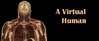
Physicians, scientists and educators around the world have now access to the most complete database of human digital images, which has allowed the development of a vast array of projects, revolutionizing the ways we understand, learn and visualize human anatomy.
 What is the Project
The Visible Human Project is a multimillion megaproject created by the National Library of Medicine (NLM), located in Bethesda, Maryland, the largest medical library in the world. Following the recommendations issued by a panel of experts in anatomy and computer-based imagery, the Project had its aim set at offering to the medical and scientific community an image database which would completely represent the body structure of a man and a woman, converted to digital format and stored into computers. Until that moment, there was no digital database of the whole human body, with all image modalities represented and taken at the same level.
NLM started then to search for the ideal human bodies that could transform the Visible Human idea into reality. The Center for Human Simulation, a renowned research center which develops advanced research projects on computerized anatomical visualization at the University of Colorado (USA), was contracted to do the systematic work of selecting among the more than two thousand cadavers donated to the Project. The fundamental condition was that the candidate body should be anatomically perfect and free of structural pathological alterations.
At the end of two long years of exhaustive search, the first "perfect cadaver" was chosen, with all organs intact: it was a 39 year old male, sentenced to death for having raped a chid, who had as only organic alterations a lethal substance injected into the circulation and the lacking of a tooth and a testicle.
This man, who had donated his body to science, has left life to be immortalized in a special paradise: the virtual medical world. Two years later, the virtual woman, who died at 59 years of age, after a sudden cardiac arrest, joined the visible man, in a remarkable gesture for the benefit of medical science and of humankind.
The Visible Human Project planned for obtaining from these cadavers four types of images: computerized x-ray tomography of the fresh and frozen body, magnetic ressonance images and anatomical cryosections. After the tomographical images were taken, each body was fully sectioned by means of a disc shaped blade, which cut the frozen tissues every 1 mm, exposing in each pass a new surface to be photographed with a high-resolution digital color camera. In the woman the slices were thinner: 0.3 mm.

In this way, the male cadaver produced 1,878 sections for each modality (frozen and fresh tomography sections, magnetic ressonance and anatomical sections) and 5,000 for the womans (also for each modality), completing a total of 19,000 images, occupying 56 Gigabytes, total (15 Gb for the man and 41 Gb for the woman).
 Applications
These images are available in the Internet, in CD-ROMs and videodiscs. Medical centers, research institutes and educational institutions are using specialized computer programs and creating tridimensional images and movies, using the Visible Human sections. There are already more than 400 projects under development in the world.
Dr. Richard Robb, from the Mayo Clinic, in Rochester, USA, was one of the first to use this database. His group developed a software called "Analyse", which is a collection of tools to processs, to display and to visualize the database in several forms. For example, this program is able to "peel" out the Visible Human, removing skin, muscle layers, etc., up to the bones, and then allowing a "virtual journey" through the remaining skeleton.

Virtual Conoloscopy: A "flight" throuhgh the human colon |
Dr Robb has also made other kinds of "flights" through the colon, the esophagus, stomach, lungs and the female reproductive system. In the future, he expects to generate virtual flights from tomographic and magnetic resonance images, in a manner similar to what is carried out with living pacients for medical diagnostic purposes. His greatest objective is to eliminate uncomfortable procedures to examine the interior of certain organs by introducing medical devices through body cavities, like endoscopes and colonoscopes. |
 There
are also virtual reality surgical simulators under development , which will
allow doctors to to train their skills and reflexes, by simulating surgical techniques before using the scapel in the patient.
Another direct impact over the patient and his doctors promoted by the Human Visible Project will be the possibility to visualize previously to the surgery the location of structures hard to be reached, like the prostate, which is surrounded by many vital organs. In this example, the doctors locate the urinary system in the screen, proceed with slicing, using the mouse as an scapel, and reach the desired system: the bladder, the prostate and urethra. Rotating the image with the mouse, they can visualize the other side of the region. This kind of visualization can be used for doctors to understand more easily anatomical structures relations among one anothers, when planning to treat or operate cancer patients.
Students are also substituting the real corpse for the virtual one.
In neuroanatomy laboratories, they admit that "the reconstructed
images give a better idea of where a variety of organs is, besides the
fact that, we are not interested only in the study of each organ in its
system, but also in the relationship with one another.
 Atlas Voxel-Man. Muscle and skin tridimensional reconstruction |
 Atlas de Voxel-Man. Muscle and skin tridimensional reconstruction with color delimitation. |
One of the programs being used by medicine students is Voxel-Man, a fascinating digital atlas developed by University of Hamburg. This atlas can be purchased by the Internet and um fascinante atlas digital desenvolvido pela Universidade de Hamburgo. Este atlas já pode ser vendido pela Internet e custa em média $200 dólares e serve como suplemento para as aulas de anatomia.
Applications develped include softwares that make tridimensional reconstruction, that allow visualization of isolated or group organs from any angle.
 Digital Humans, CD Rom available for sale |
Many softwares, CDROMs and videodiscs are already available and can be purchased even on Internet (http://www.vhd.org.br/produtos.htm). A good example is the Digital Humans CD, on which the user can "click" his mouse on any part of the Visible Human and visualize the specific section, and also make a comparison between the anatomical section and the radiological one. |
 Visible Human in Brazil
In order to easy and speedup access to other parts of the world, NLM is stablishing servers connected to the Internet in Europe, Asia and South America. According to Dr. MIchael Ackerman, NLM project coordinator, the purpose is to increase the number of institutions wiht preferential access to the image database. Until the present moment, the UNICAMP is the only institution of Latin America with projects registered with NLM.
For this reason, it granted Unicamp a image database "mirror" to be used in South America. Soon it will be installed in a 60 Gb disc computer, which will serve with this purpose only, connected to the Internet. The access is free, bur requires an access password, supplied after a project register protrocol registration with NLM, which will be intermediated by the Center for Biomedical Informatics. In case you wish to use the images, see orientations in our site (http://www.vhd.org.br/register.htm).
A demonstration containing several project images is available through WWW (World Wide Web), with text in Portuguese and free acess (http://www.vhd.org.br).
 In
Brazil, the first project using the Human Visible image database was the
Center for Biomedical Informatics (UNICAMP), through its bilingual journal
Cérebro & Mente/ Brain &
Mind, a neurosciences eletronic journal, which in one of its sections
presents brain and its structures anatomical reconstructions. In this project
, the images are still in technical improvement phase.
 Interesting Links
 The Author
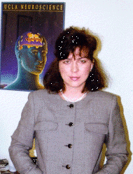
 Silvia Helena Cardoso,
PhD, psychobiologist, master degree and doctor in Sciences by the University
of São Paulo and post-doctor by the University of California, in
Los Angeles. Associate Researcher from the Center for Biomedical Informatics,
UNICAMP, Campinas, Brasil.
Email: cardoso@nib.unicamp.br
Publication
Center for Biomedical Informatics State University of Campinas © 1997 Renato M.E. Sabbatini |
Support:
Searle Farmacêutica Brasil |